




IIT Madras To Create World’s Largest 3D Human Brain Atlas Under PM Modi’s Initiative
In Short
To develop the 3D maps of over 100 human brains at the cellular resolution, PM Narendra Modi has initiated a project in the Indian Institute of Technology Madras. The project also includes C-STROKE, an open-access atlas to promote stroke damage study.

Share :
IIT Madras To Create World’s Largest 3D Human Brain Atlas Under PM Modi’s Initiative
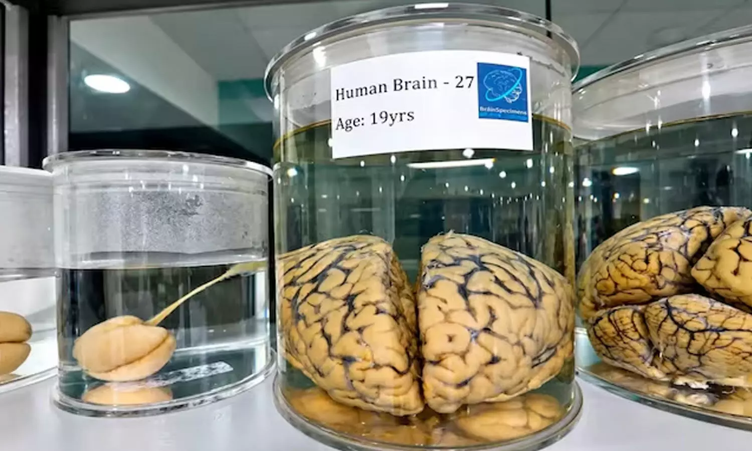
The Indian Institute of Technology Madras (IIT Madras) is in the midst of a bold initiative which seeks to map over 100 human brains in 3D, from early foetal stages to old age, including in cases of disease.
The programme which was launched by Prime Minister Narendra Modi, is aimed at putting together what the Sudha Gopalakrishnan Brain Centre (SGBC) of IIT Madras says is the world's biggest library of three-dimensional human brain maps at the cell-level.
It is hoped that the project will contribute to the understanding of how the brain develops, ages and changes during neurological diseases by researchers and medical professionals. The produced digital maps will be opened to researchers from all over the world.
IIT Madras To Map Human Brains At Cellular Resolution
The project will employ specialized imaging technology and supercomputers to transform whole human brains into detailed digital maps.
It's an effort that brings together over 200 researchers, engineers and technicians, over 10 medical institutions from India and over 30 leading researchers from more than 10 countries.
The study of the 3D structure of the brain at the cellular level could provide more insights into the structure of the brain and the changes that happen during development, throughout life and in disease.
It is being spearheaded by the Sudha Gopalakrishnan Brain Centre, which is creating cutting-edge imaging tools for neuroscience and neurological medicine.
C-STROKE Atlas provides New Insights into Stroke Damage
In his visit, Prime Minister Modi also launched C-STROKE, the world's first Human Ischaemic Stroke atlas by IIT Madras.
The atlas allows for the comparison of traditional medical imaging and microscopic analysis of the same brain that suffered a stroke. It enables scientists to examine changes in brain tissue that can be observed on clinical scans of the brain.
An ischaemic stroke is caused by a blockage in one of the blood vessels supplying the brain, depriving the brain cells of oxygen and nutrients.
Magnetic resonance imaging (MRI) is routinely used in doctors' offices to detect damage from a stroke. But MRI images can be used to see structures at the millimetre scale, whilst microscopic examination of tissue can show things at the micron scale. As a result, C-STROKE links these two levels of observation.
The atlas includes over 1,000 sections of a brain that has suffered a stroke, which are annotated. It reports neuronal health, inflammation and injury, blood-brain barrier damage, oxygen deprivation and nerve fibre damage.
The correlations between these changes in the cells and the patterns observed in MRI scans may help researchers understand more about the damage done to brain tissue by stroke and future treatment targets.
The brain atlas has been powered by Param Shakti Super Computer
The C-STROKE atlas has been developed on Param Shakti supercomputer supported by the Ministry of Electronics and Information Technology.
An open access viewer and analytical tools will allow researchers globally to explore the data and to study the microscopic changes in tissue and how this relates to the clinical imaging results.
Availability of the resource is expected to facilitate collaborative research and provide more opportunities for the study of the biological mechanisms underlying stroke.
Establishing a worldwide platform for brain research
C-STROKE is based on the brain centre's previous research such as DHARANI which was released in December 2024. This is a resource of five foetal brains with 5,132 sections.
The new project aims to expand this research over the entire lifespan by providing a more complete picture of the brain's organization across the lifespan of the individual.
The project is a combination of brain science, medical science, engineering and computing, said Prof. Mohanasankar Sivaprakasam, head of the Sudha Gopalakrishnan Brain Centre. He pointed to its possible impact on neuroscience and neurological medicine for many years to come.
By developing detailed, publicly available brain maps, IIT Madras hopes to give scientists a new tool to explore the human brain at the cellular level and deepen our understanding of neurological disorders such as stroke.


