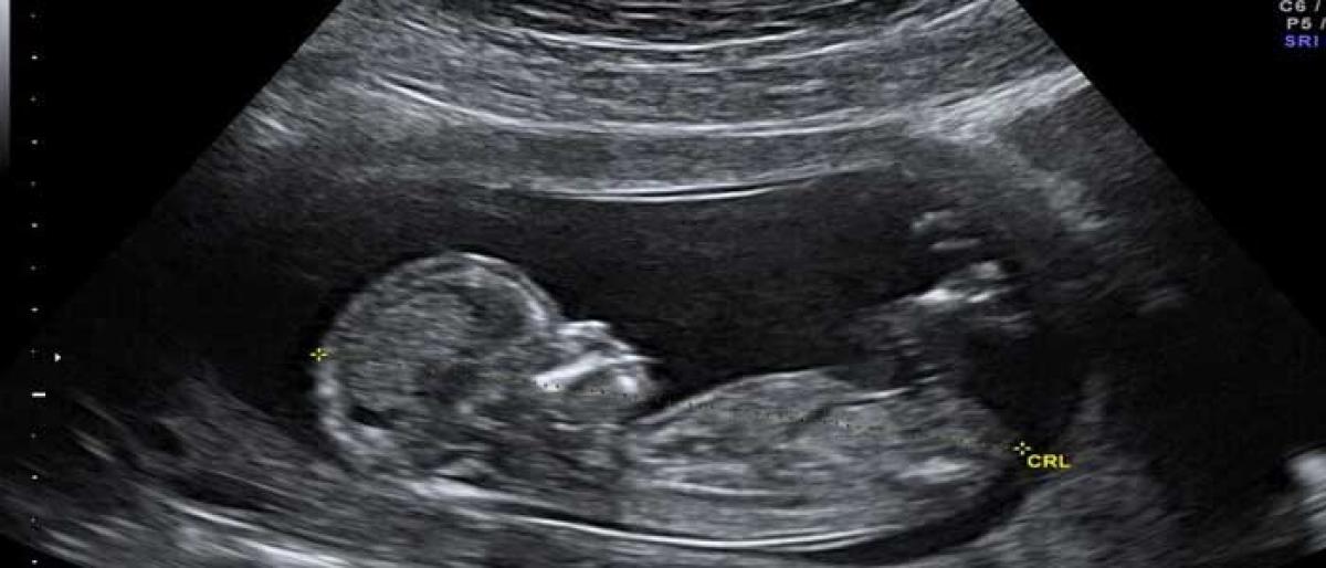
Ultrasound scan & its effect on babies

Doctors advise a mother to undergo scans during pregnancy, for both normal and high risk cases. Ultrasound scans are used more commonly today than 10 years ago and this is because scans are more accessible and affordable.
Doctors advise a mother to undergo scans during pregnancy, for both normal and high risk cases. Ultrasound scans are used more commonly today than 10 years ago and this is because scans are more accessible and affordable.
Scans are used by doctors to gather accurate information of a fetus’ growth, its position in the womb etc. throughout the pregnancy period. However, most mothers have apprehensions on the safety of these scans, whether they could harm the unborn fetus. In ultrasounds scans technology makes use of sound waves only.
The scans are to ensure if the fertilized egg has embedded itself, the baby’s heart is beating right, to detect an ectopic pregnancy (where the embryo implants outside the womb usually in the Fallopian tube), assess the risk of Down’s syndrome by measuring fluid at back of the baby’s neck at 11 to 13 weeks, check how the blood flows between your placenta and baby etc.
Ultrasound scan is vital for the gynecologists to assess the pregnancy and the infant’s development throughout the period. Doppler studies are to assess the blood flow and the fetal circulation. It can be used to evaluate growth issues as well as flow patterns in the heart, brain etc.
Fetal Echocardiography is to examine the fetal heart in detail just as an adult Echocardiogram does. Of late, 3D and 4D scans have become the rage. They give realistic images of the fetus within and helps foster the materno-fetal bond. As a diagnostic tool it is useful in certain abnormal conditions.
The pregnancy scans area to be prescribed for by the obstetrician.
Routine scans usually are:
- Dating and viability scan (between 6 and 10 weeks gestation)
- Nuchal translucency scan (also called NT/ First trimester screening scan between 11 and 13 weeks.)
- Anamoly scan/Target scan/ TIFFA scan (between 18 and 20 weeks)
- Growth scan (between 28 and 38 weeks )
More scans will be required
- In case of carrying twins or more
- If there is a difference in baby’s expected size based on the due date, and the actual of the baby measured in a scan.
- If the pregnant woman has medical complications such as high blood pressure, gestational diabetes, etc.
- Ultrasound scans can give useful and reliable information about your pregnancy. They help your gynecologist assess how your pregnancy is progressing and plan for any treatment if it is required. It has become an indispensible part of routine obstetric care. Of course, most women find scans reassuring and are simply delighted to see their baby on the screen!
By: Dr Amitha Indersen
The writer is the Head consultant Fetal medicine, Apollo Cradle hospitals, Hyderabad


