Eyes and vision

The human eye is one of the most complex and sophisticated organs in the body Its unique automatic focusing system outstrips that of any camera, and its light sensitivity is ten million times greater than the best film designed so far Before taking a look at how the eye works, lets start with a basic overview of how it is built
The human eye is one of the most complex and sophisticated organs in the body. Its unique automatic focusing system outstrips that of any camera, and its light sensitivity is ten million times greater than the best film designed so far! Before taking a look at how the eye works, let’s start with a basic overview of how it is built.
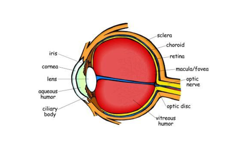
The outside layer of the eye is made up of the sclera and the cornea. The sclera is the firm white tissue that covers all of the eye except the very front. It helps maintain the shape of the eye and protects the inner parts. The cornea is the transparent portion at the center front part of the eye that allows light through. A thin outer mucous membrane called the conjunctiva covers the inside of the eyelids, the cornea, and the front portion of the sclera. It helps lubricate the eye.
The middle layer of the eye contains oxygen- and nutrient-rich blood vessels, most of which are located in the layer of tissue called the choroid. Near the front of the eye is the ciliary body, a group of muscles and ligaments that attach to the lens. These muscles change the shape of the lens as they relax and contract. The final component of this layer is the iris, a group of muscles that controls how much light enters the eye by adjusting the opening, or pupil.
The iris contains pigments that determine your eye-color. When you look at a person’s eye, you can see parts from each of the first two layers: the “white” of the eye is the sclera, the front transparent part is the cornea, the iris is the colored part, and the pupil is the dark hole in the center.
The eye’s inner layer is composed of the retina: thin tissue that contains blood vessels and light-sensitive photoreceptor cells called rods and cones. Every human eye contains about 120 million rods and 7 million cones. The rods are very sensitive to low-level light, but they cannot distinguish color. Cones need much more light to function than rods do, but they provide color information and sharp detail. You may have noticed that in dim light color looks much less vibrant; that is because the rods that help you see in the dark are more or less “color-blind.” The retina also contains a dark pigment called melanin (also found in skin and hair cells) – this reduces reflection of light when it enters your eye.
The blood vessels and the optic nerve(the nerve that conducts electrical impulses to the brain; see the following article to learn more) connect to the retina at a spot called the optic disc. On this disc there are no rods and cones; this is your blind spot. You don’t usually notice your blind spot, because your two eyes work together to “cover up” each other’s blind spot.
The macula is a small spot in the center of your retina. On this spot is a small pit called the fovea. When light is focused on this spot we get the sharpest image, because the fovea contains very tightly-packed photoreceptor cells. Macular degeneration is a common eye disease that is caused by the deterioration of the macula and results in partial blindness.
The three layers fill only a small part of the eye; the large middle area isn’t empty, though! The area between the cornea and the lens is filled with a transparent liquid material called the aqueous humor.The area between the lens and the retina contains a clear gel-like substance called the vitreous humor. Both of these humors help give shape to the eye and are part of the focusing process. Your eye is a very delicate organ. The sclera and cornea protect the inner parts of the eye, but there are other protective parts as well.


